this post was submitted on 13 May 2024
151 points (99.3% liked)
Ask Science
8356 readers
1 users here now
Ask a science question, get a science answer.
Community Rules
Rule 1: Be respectful and inclusive.
Treat others with respect, and maintain a positive atmosphere.
Rule 2: No harassment, hate speech, bigotry, or trolling.
Avoid any form of harassment, hate speech, bigotry, or offensive behavior.
Rule 3: Engage in constructive discussions.
Contribute to meaningful and constructive discussions that enhance scientific understanding.
Rule 4: No AI-generated answers.
Strictly prohibit the use of AI-generated answers. Providing answers generated by AI systems is not allowed and may result in a ban.
Rule 5: Follow guidelines and moderators' instructions.
Adhere to community guidelines and comply with instructions given by moderators.
Rule 6: Use appropriate language and tone.
Communicate using suitable language and maintain a professional and respectful tone.
Rule 7: Report violations.
Report any violations of the community rules to the moderators for appropriate action.
Rule 8: Foster a continuous learning environment.
Encourage a continuous learning environment where members can share knowledge and engage in scientific discussions.
Rule 9: Source required for answers.
Provide credible sources for answers. Failure to include a source may result in the removal of the answer to ensure information reliability.
By adhering to these rules, we create a welcoming and informative environment where science-related questions receive accurate and credible answers. Thank you for your cooperation in making the Ask Science community a valuable resource for scientific knowledge.
We retain the discretion to modify the rules as we deem necessary.
founded 1 year ago
MODERATORS
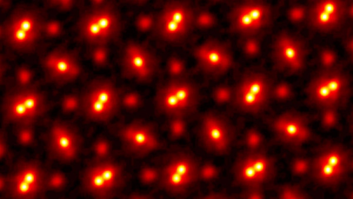
Each of the orbs are the atoms. The brighter orbs that are nearest each other at the Praseodymium (Pr) atoms. The single orbs are the Scandium (Sc) atoms and the less prominent orbs are the Oxygen atoms. I believe based on the content of the article.The space between is just that, the space between the atoms. They are all in a lattice pattern due to how they are attracted to each other.
I have a followup question: When scientists are taking these images, are they just static snapshots or does the technology to see them doing their thing in real time exist? Are they even actually "pictures" in that sense, or are they just representations of things we can't actually see?
These images are generated from processing many out of focus images while scanning across the area. They use the differences between the out of focus images to compute what must have caused those differences.
So this technique is pretty far from being able to capture real time events as it requires capturing hundreds of images to produce a single computed image. The paper talks about how thermal motion of the nuclei is what now dominates the limits of resolution for this method.
There are other ultrafast imaging techniques that can capture essentially real-time chemical reactions, but I believe those don't come close to this spatial resolution.
Not 100% comparable, but synchrotron XRD allows for real-imaging of solid state chemical reactions and can, in a sense, resolve the unit cell structure of the crystal. However, what you get from an XRD is nothing like this "photo-like" image, but a diffractogram. I think you could probably re-create an image like this from a 2D diffractogram though, but I'm not sure.
I could be wrong, but I think XRD requires very pure crystals of sufficiently large size. That can help you ascertain the structure and composition of something you can synthesize and crystalize, but I don't believe xrd can image specific regions of interest like single doping sites like this article talks about.
Synchrotron XRD also has a major drawback in having a very significant equipment requirement that requires being able send samples away for analysis at a dedicated facility. That puts limits on sample preparation and stability time, as well as sharing beam time with lots of other groups.
It's been a while since I've read up on synchrotron xray diffraction though, so there could be workarounds for some of those limitation or I could be misremembering details.
You're definitely correct that getting sychrotron time is hard :(
On the first part though: Yes and no. XRD will tell you about things like strain and unit cell size distribution, so in that sense, you can't resolve a single doping site. On the other hand, if you have a reaction going on, or some dopant diffusing into your sample, synchrotron XRD is powerful/fast enough that you can "film" how the crystal structure changes in real time. That "film" will be a kind of average of many sites, but can still be focused to a relatively small region (don't remember exactly how small off the top of my head, but I believe we're talking nm-scale).